
")
")


 |
 |
 |
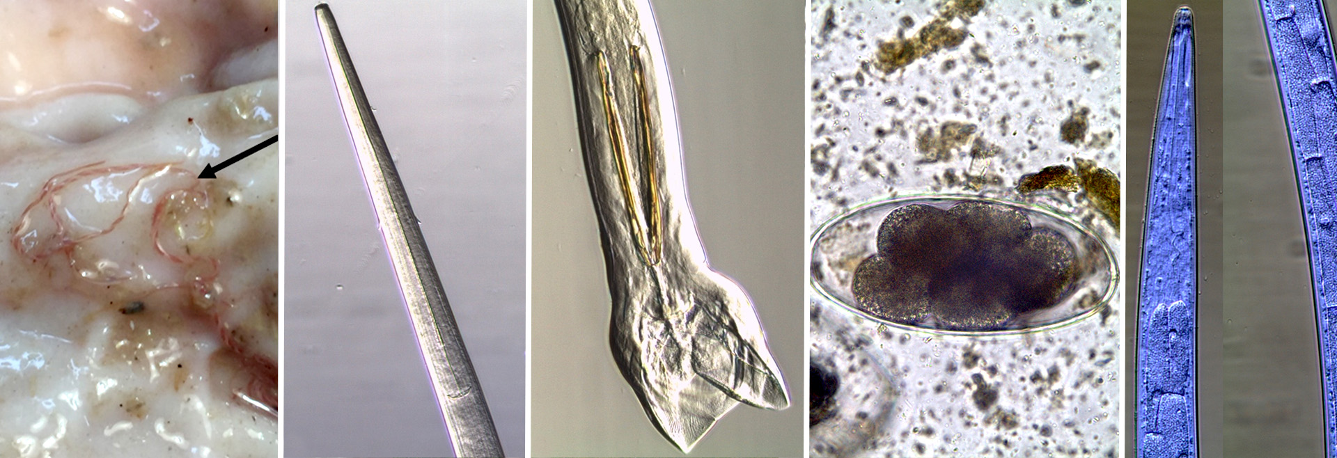
Fasciolosis is a liver parasitosis caused by trematodes of the genus Fasciola.
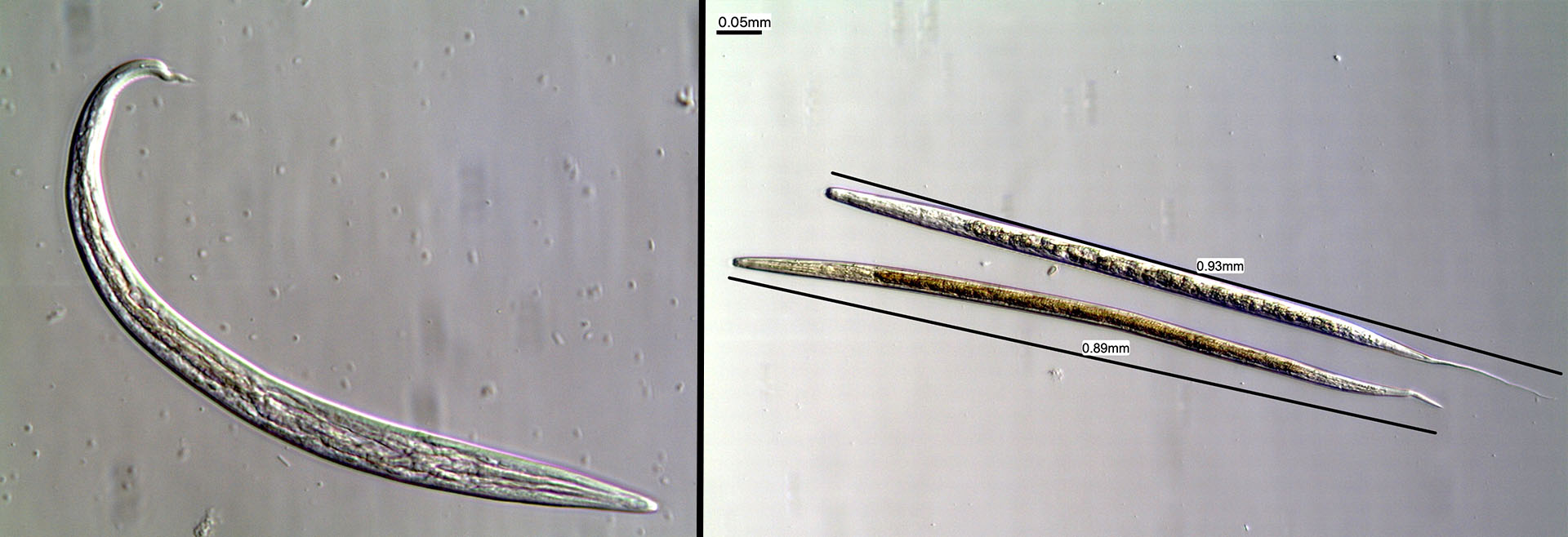
In Portugal, the species present is Fasciola hepatica. It is one of the largest trematodes in the world and can measure up to 30 mm long by 13 mm wide. The development of F. hepatica involves a complex life cycle, which requires an intermediate mollusc host of the Lymnaeidae family, usually Galba truncatula, to be completed. Definitive hosts and intermediate hosts must exist in the same place for infection to be established.
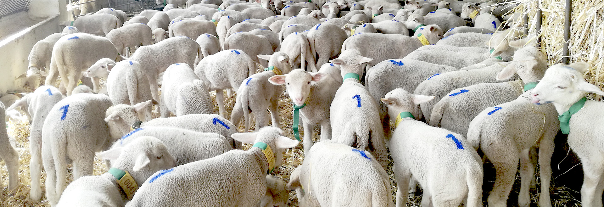
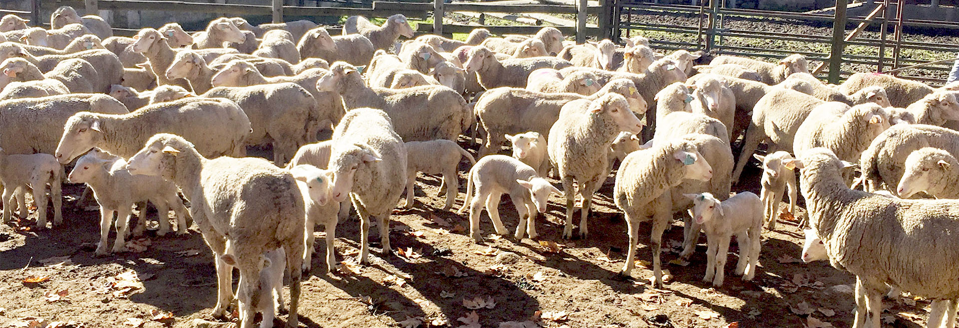
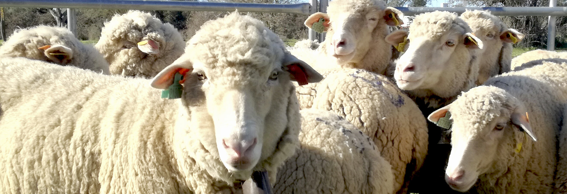
This parasite infects a wide range of hosts, including humans, as it is a zoonotic agent. Small ruminants are especially sensitive and may become reinfected several times during their lifetime since they do not develop immunity to this parasitosis.
Depending on the parasite load, clinical signs include anaemia, abdominal distension, and sudden death during the migration phase of the larval forms through the liver parenchyma (acute phase), and anaemia, weakness, submandibular oedema, and decreased milk production due to the presence of adult parasites in the bile ducts (chronic phase).
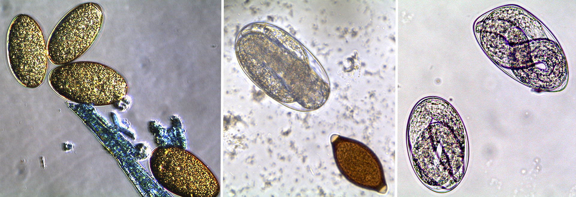
The eggs produced by the adults are first transported into the intestinal lumen with the bile and then to the outside with the faeces where they can be detected using coprological techniques, such as the sedimentation technique.
The infection also results in the production of antibodies that can be tested for using serological techniques. The sedimentation technique only detects chronic infections, whereas serological techniques allow diagnosing both the early stages of infection, before there is egg production, and chronic infections, even when egg production is low.
Antibodies are no longer detectable 6 to 12 months after parasite clearance.
The blue bar represents the percentage of positive samples where antibodies against F. hepatica were detected, and the orange bar represents the percentage of negative samples.
The blue bar represents the percentage of stool samples positive for eggs and the orange bar represents the percentage of negative samples.
The box-plot shows only the results obtained for the White Merino sheep because no eggs were detected in the faeces of the Black Merino sheep. The horizontal line dividing the box indicates the median value of eggs per gram of faeces (OPG) observed, the lower line the minimum value and the upper line the maximum value. Outliers are represented by a circle.